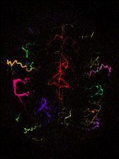
Vascular Territory Map

Dynamic angiography above the circle of Willis

Combined angiography and perfusion imaging: angiographic reconstruction

Combined angiography and perfusion imaging: perfusion reconstruction

Thomas Okell
Associate Professor
- Sir Henry Dale Fellow
- Head of Neurovascular Imaging
- OxCIN MRI Graduate Programme Director
My research focusses on the development of novel non-invasive MRI methods which visualise blood flowing through the arteries that feed the brain and the resulting perfusion of the brain tissue. Much of my initial research focussed on developing techniques which allow blood from individual feeding arteries to be followed through the vascular tree. In addition to providing information about the structural and functional status of each artery, these methods allow the assessment of "collateral blood flow". This is when the main feeding artery to a certain brain region becomes blocked or significantly narrowed, but the flow of blood from secondary arteries maintains perfusion in this brain region, preventing a significant stroke. The presence or absence of collateral flow can be important in deciding between potential treatment options in patients with arterial disease. These vessel-selective strategies also have applications in diseases where the arterial source of blood flow is important, such as tumours and arteriovenous malformations.
In my previous five year research fellowship from the Royal Academy of Engineering, I aimed to address one of the key downsides to these imaging techniques, which is that obtaining 3D time-resolved images of the arteries as well as maps of tissue perfusion is time-consuming, and therefore difficult to apply in a clinical setting. I designed a single scan which can track the flow of blood through the arteries, all the way into the tissue, thereby providing both sets of information at the same time. I used recently developed acquisition and image reconstruction methods to accelerate this process, allowing images to be acquired in a fraction of the time normally required.
My current Sir Henry Dale Fellowship, jointly funded by the Wellcome Trust and the Royal Society, aims to develop new brain blood flow imaging methods using a powerful ultra-high field MRI scanner. There are a series of technical challenges to solve to make these methods efficient and robust, but once these have been overcome, highly sensitive measurements of brain blood flow will be possible. I plan to use this improved sensitivity to obtain very high spatial and temporal resolution information, as well as examine blood flow to the white matter of the brain, which is extremely challenging using conventional scanners, but has relevance to a broad range of conditions, including dementia.
I will continue to trial existing techniques and new methods, as they emerge, in collaboration with clinical colleagues in a range of patient groups, including those with stroke, arteriovenous malformation and vascular cognitive impairment. I hope that this will show the potential utility of these techniques for understanding the progression of these diseases, and in the longer term help with diagnosis, prognosis and therapeutic planning in these patients.
Research groups
Team Members and Collaborators
-
Hao Li
DPhil Student
-
Qijia Shen
DPhil Student
-
Joseph Woods
Postdoctoral Researcher in Ultra-High Field MRI
-
Yuriko Suzuki
Royal Academy of Engineering Research Fellow
-
Peter Jezzard
Herbert Dunhill Professor of Neuroimaging
Key publications
-
Dynamic B0 field shimming for improving pseudo-continuous arterial spin labeling at 7 Tesla
Journal article
Ji Y. et al, (2024), Magnetic Resonance in Medicine
-
Multimodal population brain imaging in the UK Biobank prospective epidemiological study
Journal article
Miller KL. et al, (2016), Nature Neuroscience, 19, 1523 - 1536
-
The dorsal posterior insula subserves a fundamental role in human pain
Journal article
Segerdahl AR. et al, (2015), Nature Neuroscience, 18, 499 - 500
-
Combined angiography and perfusion using radial imaging and arterial spin labeling
Journal article
Okell TW., (2019), Magnetic Resonance in Medicine, 81, 182 - 194
-
A general framework for optimizing arterial spin labeling MRI experiments
Journal article
Woods JG. et al, (2019), Magnetic Resonance in Medicine, 81, 2474 - 2488
-
Cerebral Blood Flow Quantification Using Vessel-Encoded Arterial Spin Labeling
Journal article
Okell TW. et al, (2013), Journal of Cerebral Blood Flow & Metabolism, 33, 1716 - 1724
-
Vessel-encoded dynamic magnetic resonance angiography using arterial spin labeling
Journal article
Okell TW. et al, (2010), Magnetic Resonance in Medicine, 64, 698 - 706
-
Recent technical developments in ASL: A Review of the State of the Art
Journal article
Hernandez-Garcia L. et al, (2022), Magnetic Resonance in Medicine
Recent publications
-
Brain Perfusion Imaging of a Large Population: Arterial Spin Labelling MRI in UK Biobank
Preprint
Okell TW. et al, (2025)
-
MOdulation-Guided ENcoding (MOGEN) Scheme for Vessel-Encoded Arterial Spin Labeling
Preprint
Li H. et al, (2025)
-
Achieving robust labeling above the circle of Willis with vessel‐encoded arterial spin labeling
Journal article
Li H. et al, (2025), Magnetic Resonance in Medicine
-
SNR‐efficient whole‐brain pseudo‐continuous arterial spin labeling perfusion imaging at 7 T
Journal article
Woods JG. et al, (2025), Magnetic Resonance in Medicine
-
Combined Angiographic, Structural and Perfusion Radial Imaging using Arterial Spin Labeling
Preprint
Okell TW. et al, (2025)
-
Ultra‐high temporal resolution 4D angiography using arterial spin labeling with subspace reconstruction
Journal article
Shen Q. et al, (2025), Magnetic Resonance in Medicine, 93, 1924 - 1941
-
Few-shot learning for highly accelerated 3D time-of-flight MRA reconstruction
Preprint
Li H. et al, (2025)
-
SNR-Efficient Whole-Brain Pseudo-Continuous Arterial Spin Labeling Perfusion Imaging at 7 Tesla
Preprint
Woods JG. et al, (2024)
-
Dynamic B0 field shimming for improving pseudo-continuous arterial spin labeling at 7 Tesla
Journal article
Ji Y. et al, (2024), Magnetic Resonance in Medicine
-
Trial of the cerebral perfusion response to sodium nitrite infusion in patients with acute subarachnoid haemorrhage using arterial spin labelling MRI.
Journal article
Ezra M. et al, (2024), Nitric Oxide, 153, 50 - 60


